


Eyelid surgery training models
3D printed, multi-layer eyelid and strabismus models on which ophthalmic, oculoplastic and plastic surgeons practise blepharoplasty, ptosis, ectropion, canthal and muscle surgery before they operate on a patient.
Marking the incision on an earlier revision of the lid model at a LidSUMMIT station.
- 3 modelseyelid, lateral canthus, extraocular muscles
- 5 layersskin, connecting tissue, muscles, fat, bone
- 50+ volumesfrom 7 base materials per lid model
- Made in Austriaby eyecre.at GmbH, Innsbruck
Three models, each for its own part of the surgery
Every model is ordered and shipped by eyecre.at. Prices as in our shop, excluding VAT and shipping, minimum order 1 unit.
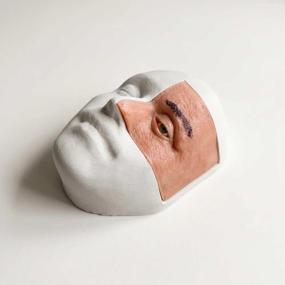
M.A.R.K.O. LID
The full multi-layer eyelid with brow, lid margin and lateral orbital rim. Our most comprehensive oculoplastic model, refined course by course at the LidSUMMIT.
- Upper and lower blepharoplasty
- Ptosis correction, including frontalis sling
- Ectropion repair with lateral tarsal strip
- Canthoplasty and canthopexy
- Eyelid malposition correction
- Direct, advancement and rotation flaps
- Wound drainage simulation and closure
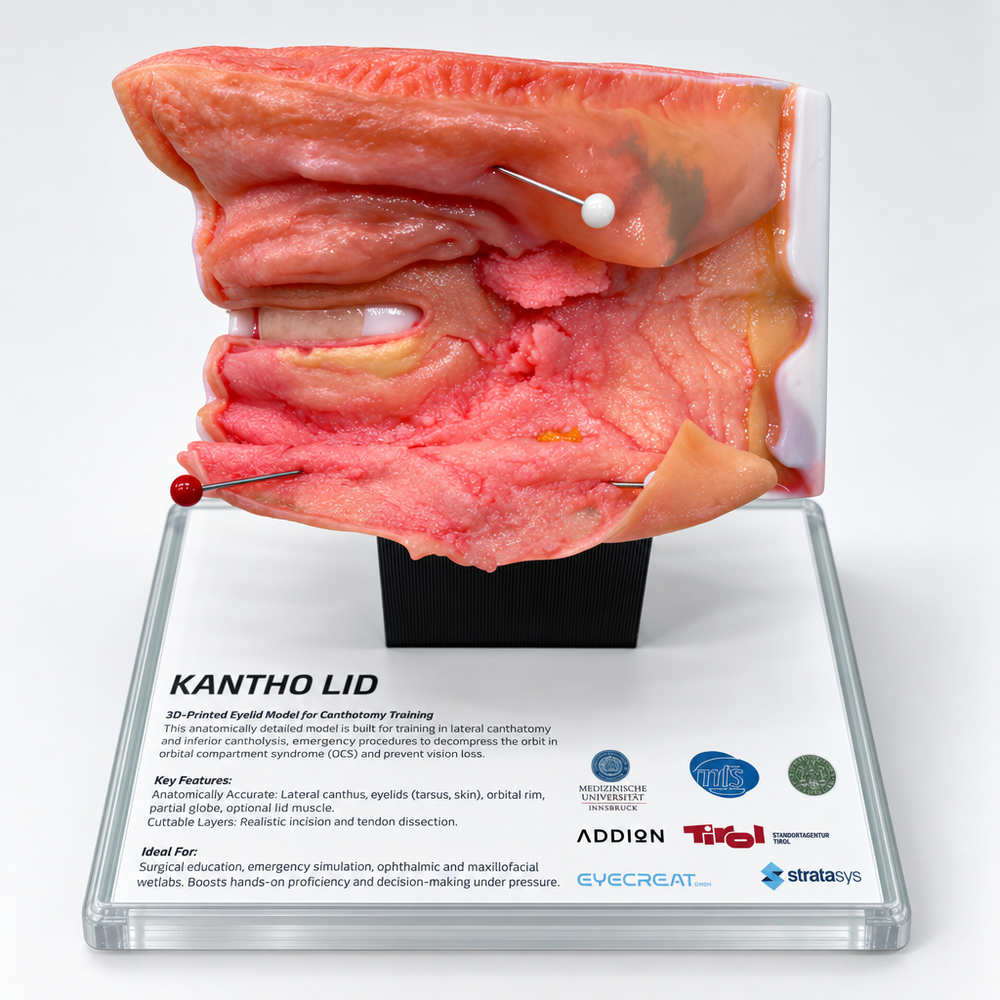
KANTHO LID
Focused on the lateral canthus: the time-critical canthotomy and cantholysis, and canthal repair afterwards. For fellowships, emergency medicine wetlabs and congresses.
- Lateral canthotomy
- Inferior and superior cantholysis
- Canthoplasty and lateral canthal repair
- Trauma-related eyelid wound management
- Emergency orbital decompression simulation
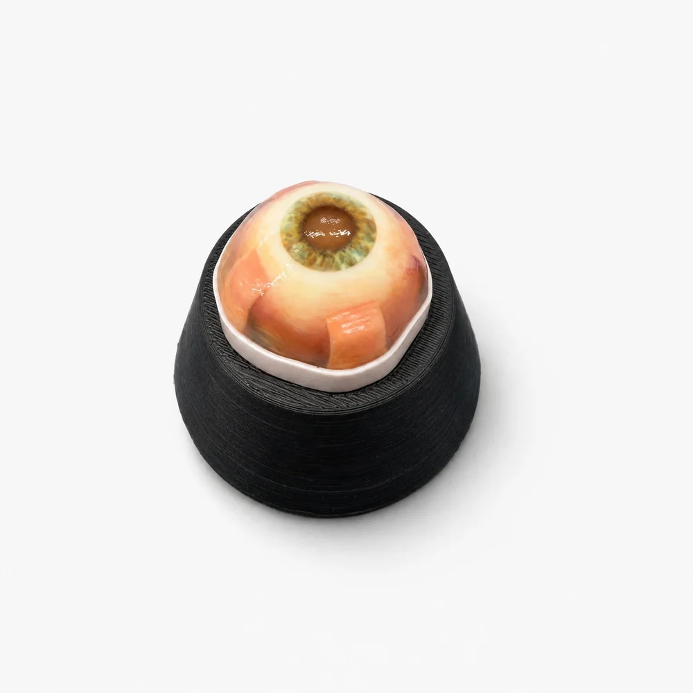
Eye 4 Strabismus
Globe with cornea and sclera, four rectus muscles and a conjunctiva over the insertions, in a frame. Eye only, or with the Holder 4 Strabismus (PN0458 plus PN0495) for 358 euros.
- Conjunctival opening and closure
- Isolating and hooking the rectus muscles
- Muscle recession and resection
- Suture placement at the muscle insertion
- Paediatric strabismus surgery familiarisation
Planning a course with many stations? Ask for a course set: models, heads, sutures and instruments in the quantities you need, with volume pricing.
Head, sutures and instruments
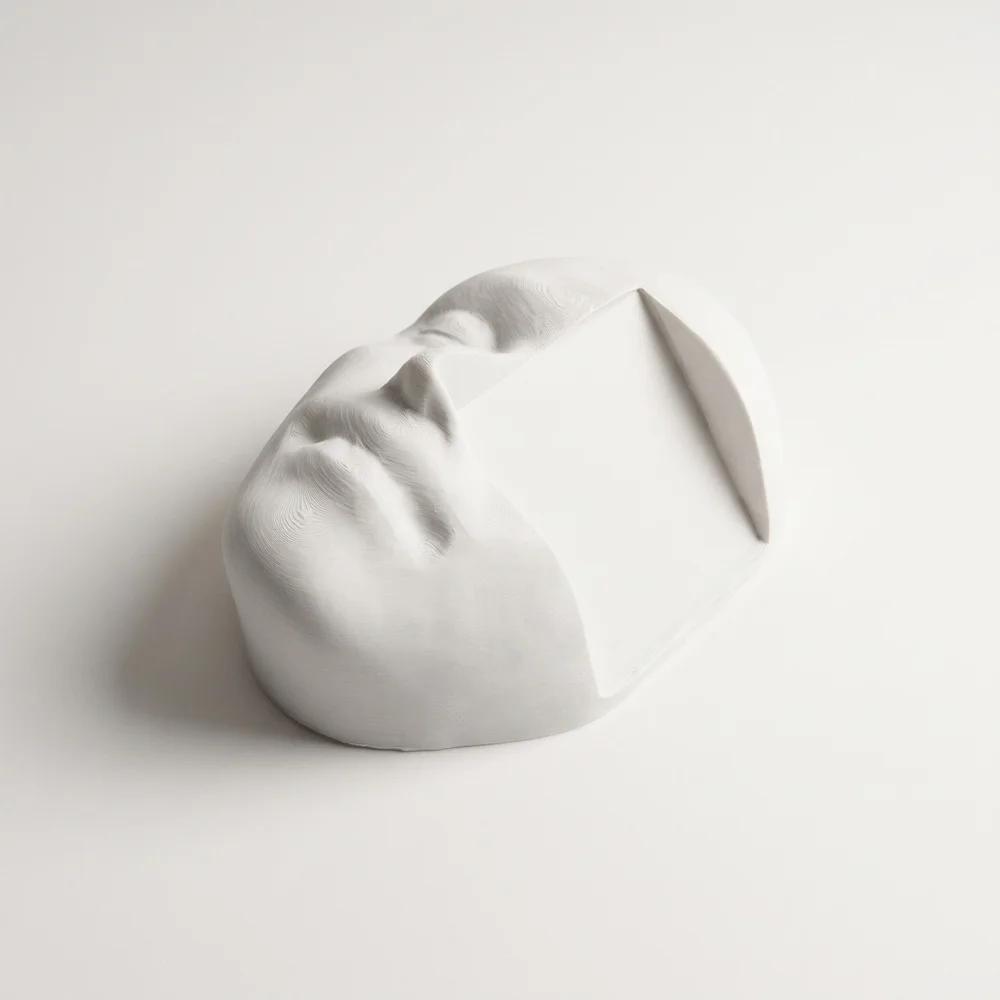
Head 4 Eyelid
Training head that holds the M.A.R.K.O. LID and KANTHO LID in a natural position at the table. €190, Art. no. PN0443.
Head 4 Eyelid in the shop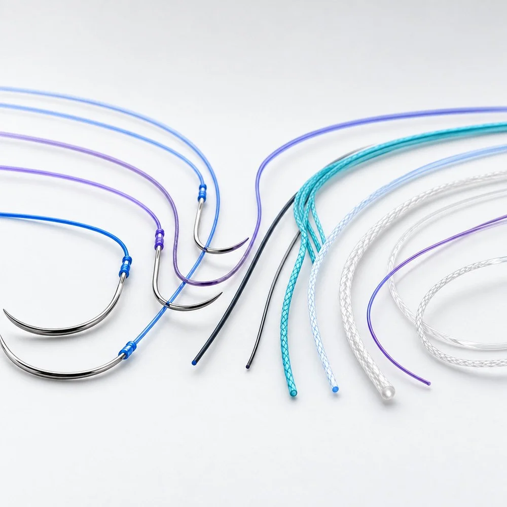
Sutures 4 Practice
For both lid models we recommend the Suture 5/0 Polypropylene Blue 90 cm Double (PN0462, €5), for the Eye 4 Strabismus the 6/0 Polypropylene Blue 45 cm Single (PN0466, €9). Range from €3.
Sutures 4 Practice in the shop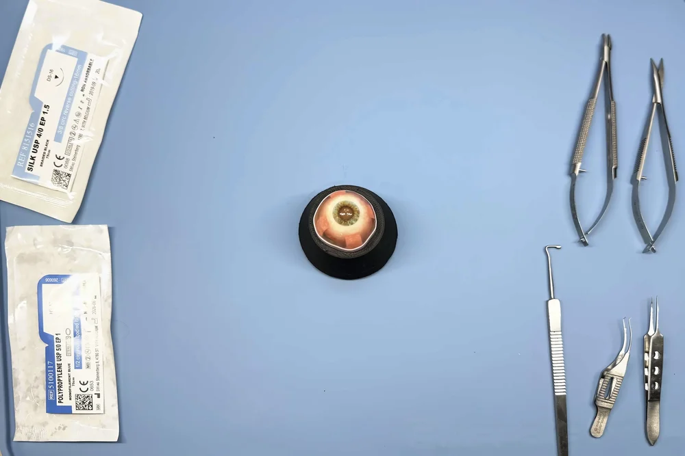
Instruments and knives
Practice instruments for lid and muscle surgery, sold individually: needle holders, forceps, scissors, muscle hooks, lid retractors and more.
Instruments 4 Practice · Knives 4 PracticeHow the M.A.R.K.O. LID is built
M.A.R.K.O. stands for Multi-Layer Anatomical Reconstructive Kit for Oculoplastics. The lid is printed layer by layer, so every layer behaves differently under the blade, as it does in a real lid.
Five anatomical layers
- Skin
Material with flow fibres for realistic cutting and suturing in blepharoplasty. - Connecting tissue
The orbital septum follows the skin closely. - Muscles
Frontalis close to the bone, with space to the skin for frontalis sling training. - Fat
Realistic fat tissue behind the septum. - Bone
Flat frontal bone and periosteum on the lateral orbital rim for anchoring the tarsus under tension.
| Volumes | More than 50 separately defined volumes, each with its own material recipe |
|---|---|
| Materials | 7 base materials, blended into Stratasys digital materials during the print |
| Printer | Multi-material PolyJet printing on a Stratasys J850 Digital Anatomy |
| Finishing | Post-processed by hand with several chemical treatments |
| Blood | Contains artificial blood; store airtight so it stays liquid |
| Scope | Lacrimal canaliculi indicated but not functional; nasal region and maxillary and nasal bones schematic only |

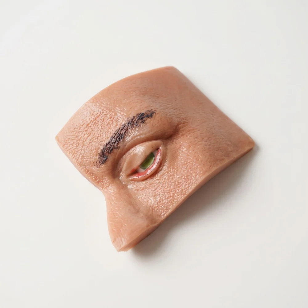

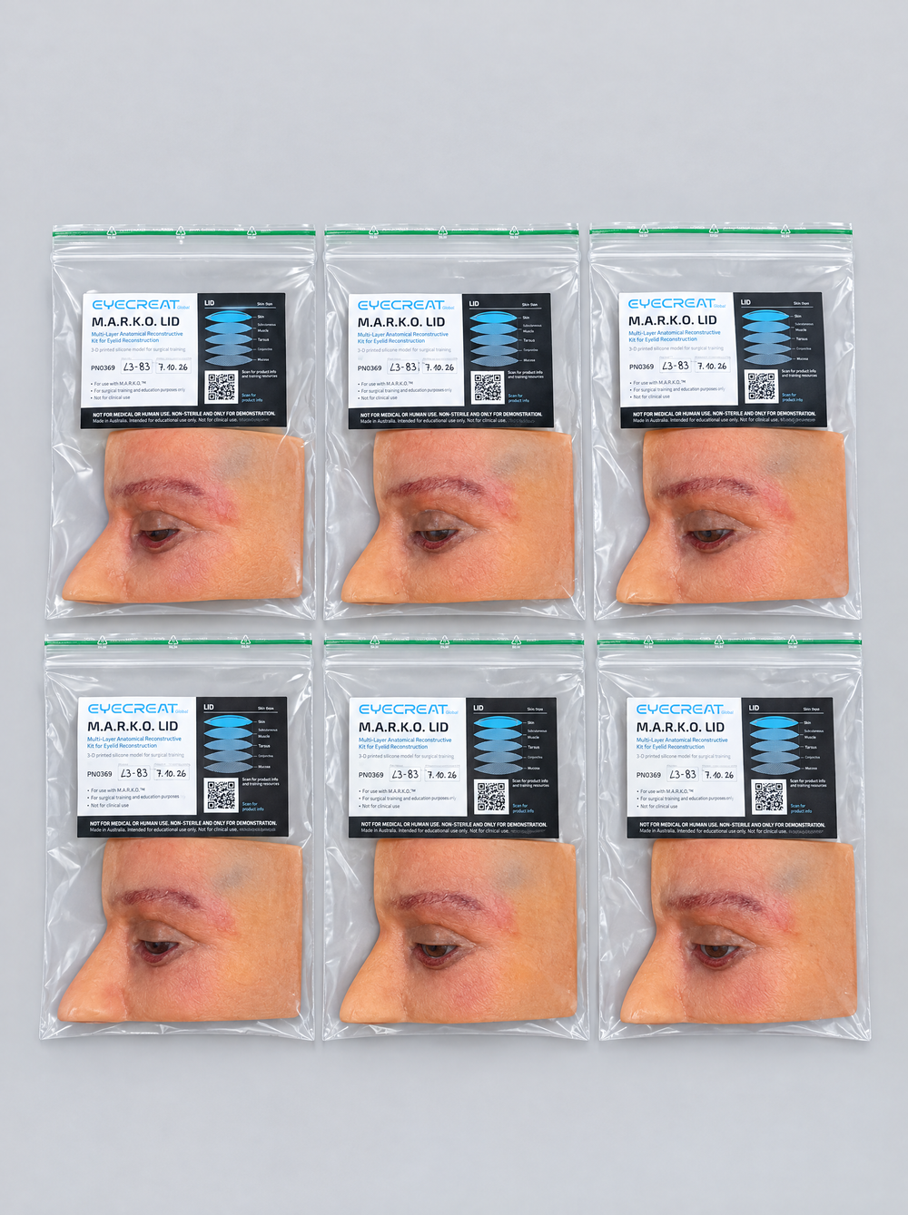
Left to right: the lid insert, a close view of the skin and lid margin (AI-enhanced video still), six inserts packed in resealable bags.
- Ectropion simulation: the lower lid everts outward for lateral tarsal strip surgery
- Natural proportions between medial lid margin, nasal bridge, eye and brow
- Skin sits loosely on the frame, giving a tension-free model
- Current revision L3-B1, with a new frame and head


It took a whole community to build an eyelid
The idea came up with Priv.-Doz. Dr. Jens Heichel and Prof. Dr. Arne Viestenz from University Medicine Halle. It produced the Eyelid 4 Eye and the KANTHO LID, and then stalled: a full eyelid needed funding and anatomical and technical know-how that we did not have alone.
It became real through a funded cooperation with the Institute of Clinical and Functional Anatomy of the Medical University of Innsbruck, led by Univ.-Prof. Dr. Marko Konschake, and with Addion, which brought multi-material printing on the Stratasys J850 Digital Anatomy. The project was funded by the State of Tyrol through the Tiroler Innovationsförderung.
- Before 2022First idea in Halle. Eyelid 4 Eye and KANTHO LID.
- 2022 to 2024Funded project of eyecre.at, Addion and the Medical University of Innsbruck.
- Since 2025Used at LidSUMMIT next to human specimens, improved after every course.
- TodayRevision L3-B1. Development continues.

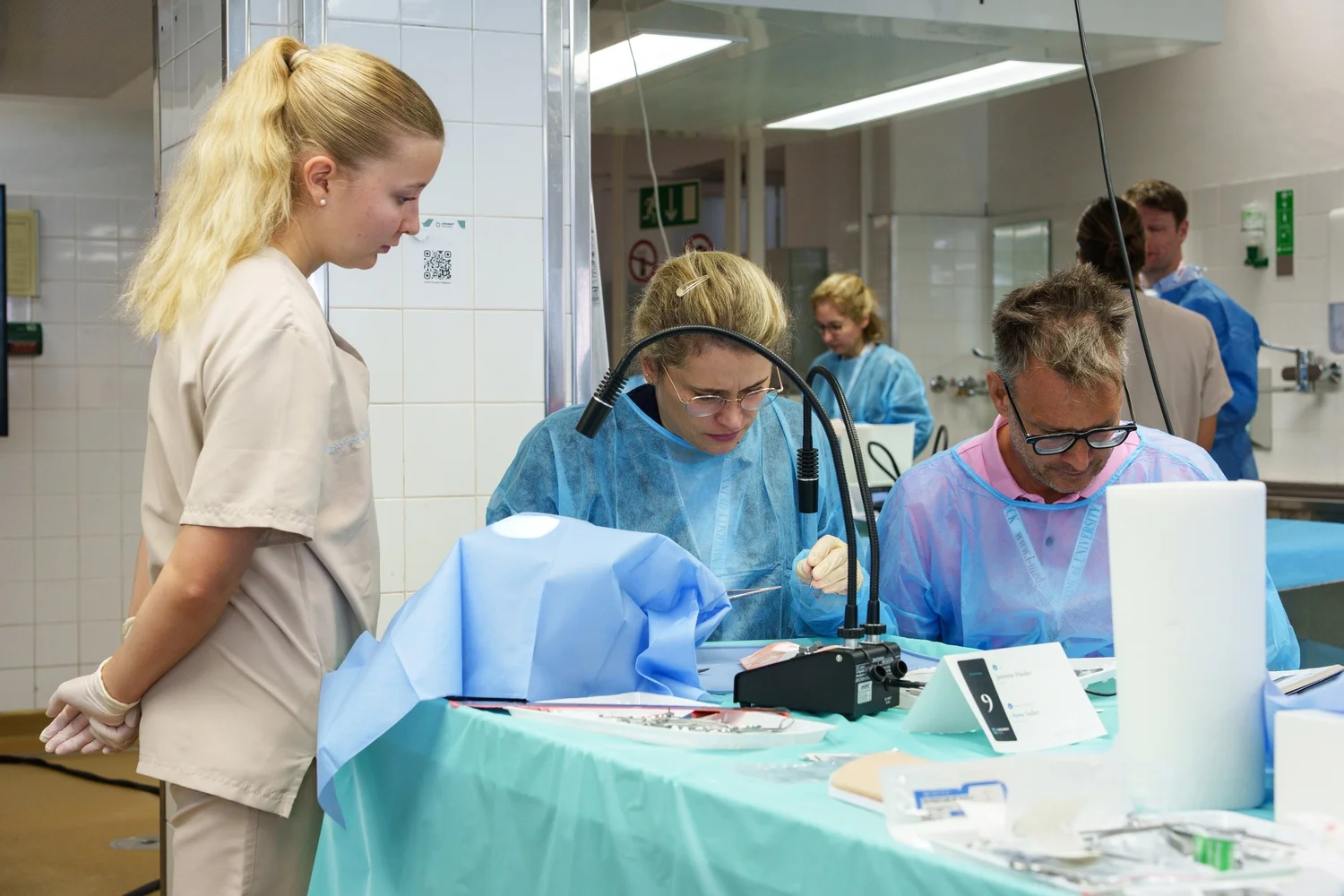
LidSUMMIT: tested and improved, course by course
The LidSUMMIT is our hands-on course for eyelid surgery at the Medical University of Innsbruck. Surgeons train on the M.A.R.K.O. LID next to human specimens, and what faculty and participants tell us goes straight into the next revision of the model.
- Next edition
- 15 to 17 March 2027
- Where
- Innsbruck
- Seats
- Max. 40, two per station
Photo: participants at their station during the LidSUMMIT.
Frequently asked questions
Which procedures can I train on these models?
On the M.A.R.K.O. LID: upper and lower blepharoplasty, ptosis correction including frontalis sling, ectropion repair with lateral tarsal strip, canthoplasty, canthopexy, malposition correction, flap reconstruction and wound closure. On the KANTHO LID: lateral canthotomy, cantholysis and canthal repair. On the Eye 4 Strabismus: conjunctival opening, muscle hooking, recession and resection.
How should I store the models?
Keep each model in an airtight container or a ziplock bag between sessions. This keeps the artificial blood inside the model liquid. Store the models at room temperature and out of direct sunlight, because cold makes the material harder.
Can a model be used more than once?
Yes, for several training rounds, as long as you work on areas that are still intact. Tissue that has been cut does not grow back, so the number of rounds depends on the procedures you practise. We recommend the 5/0 polypropylene suture (PN0462) because it tears out less quickly in the model's layers, which gives you more rounds per model.
Do you offer course sets, volume pricing or custom versions?
Yes. For residency programmes, wetlabs and congresses we put together course sets with models, heads, sutures and instruments, and quote volume prices. We also adapt existing models and develop new ones with you; for small adaptations the first prototype typically takes 1 to 3 months. Use the request form on this page.
How long does delivery take?
Our models are made to order in Innsbruck, Austria. Production usually takes about 2 weeks, then UPS delivers to most countries within 1 to 5 business days. We ship worldwide. If you need the models for a fixed course date, tell us when you order.
Are the models medical devices?
No. All models are training and demonstration aids: NOT FOR MEDICAL OR HUMAN USE, NON-STERILE AND ONLY FOR DEMONSTRATION.
Request a quote or a course set
Tell us what you want to train and for how many surgeons. Our team in Innsbruck answers every request personally.
- Single models for you or your department
- Course sets and volume requests for wetlabs and congresses
- Custom adaptations and new models
- Questions about the LidSUMMIT

Prefer e-mail? Write to request@eyecre.at or call +43 699 150 97 860.