Vitrectomy Wetlab Training: A Practical Guide to Practising PPV
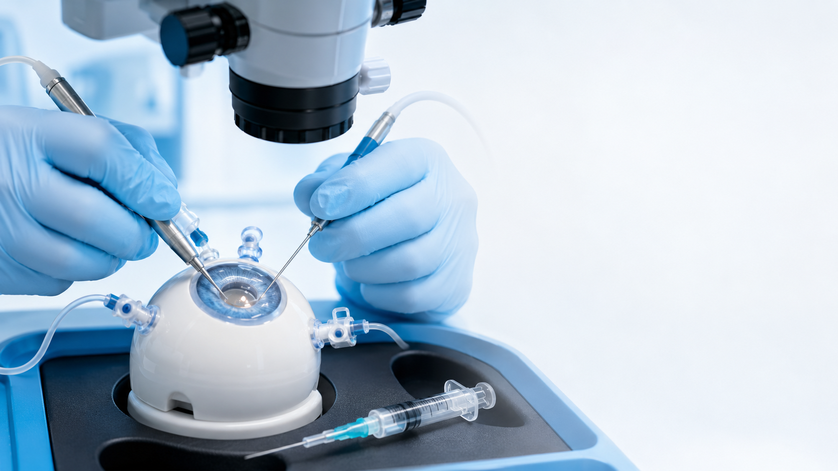
Pars plana vitrectomy (PPV) is one of the most technically demanding procedures in ophthalmology. Trocar placement, core vitrectomy, membrane peeling, endoillumination, fluid-air exchange and tamponade each carry their own learning curve, and none of them are skills you want to learn for the first time in theatre. A well-built wetlab lets a surgeon repeat every one of those steps until the hands know them.
This guide walks through the PPV procedure step by step and shows exactly which training model, vitreous substitute and accessory supports each stage, so you can put together a vitrectomy wetlab that mirrors the real operation.
What is a pars plana vitrectomy?
A vitrectomy removes the vitreous gel from the posterior segment of the eye. Access is gained through the pars plana, roughly 3.5 to 4 mm behind the limbus, where instruments can be introduced without damaging the retina or the lens. Once the vitreous is removed, the surgeon can address the underlying pathology: retinal detachment, epiretinal membrane, macular hole, vitreous haemorrhage, diabetic tractional disease and more.
Modern PPV is a multi-port, sutureless technique. The surgeon works with a vitrectomy cutter, an endoillumination probe and an infusion line simultaneously, all under microscope visualisation. Every instrument exchange and every fluid manoeuvre has to be precise, which is exactly why repetition-based wetlab practice matters so much.
For a full clinical overview of the procedure, indications and instrumentation, see the American Academy of Ophthalmology's reference article: Pars Plana Vitrectomy on EyeWiki.
Why practise vitrectomy in a wetlab?
Vitreoretinal training has traditionally leaned on porcine eyes and expensive virtual simulators. Both have limits: porcine tissue is inconsistent, perishable and never quite matches human anatomy, while high-end simulators are costly and rarely available outside a handful of centres.
A dedicated artificial training eye sits between the two. It gives consistent, repeatable anatomy that behaves the same way every time, it can be set up on any bench, and it lets a resident run the same manoeuvre twenty times in an afternoon. For a course organiser it means every station is identical, so every participant gets the same experience.
The PPV procedure, mapped to your wetlab
Here is each core step of a vitrectomy and the eyecre.at equipment that lets you train it.
1. Port placement and trocar insertion
The first skill is confident, correctly angled trocar placement through the pars plana. The Eye 4 Vitrectomy model (AS0052) is engineered for exactly this, with the anatomy needed to practise 20-, 23-, 25- and 27-gauge port creation and instrument introduction. Pair it with 20G trocars and infusion line to build the full three-port setup.
2. Core vitrectomy
With ports in place, the surgeon removes the central vitreous. This is where a realistic vitreous substitute is essential. The Vitreous Body Gel is a synthetic vitreous humour substitute that behaves like the real gel under the cutter, available in 500 ml, 1000 ml and 2000 ml volumes so a whole course can be supplied from a single order.
3. Visualisation and membrane peeling
Peeling epiretinal and internal limiting membranes depends entirely on seeing them. Dye for Vitreous (a triamcinolone-style visualisation solution) makes the vitreous and membranes visible, so trainees can practise staining and controlled peeling technique. For background on the clinical technique, the AAO reference is Epiretinal Membrane on EyeWiki.
4. Endoillumination
Vitreoretinal work happens in a darkened field lit only by the endoillumination probe. The Illumination 4 Eye accessory (AS0088) adds realistic internal illumination to the training eye, so surgeons get used to working with the light probe in one hand and the cutter in the other.
5. Retinal manipulation and heavy liquid
For detachment cases, perfluorocarbon liquid is used to flatten and reposition the retina. Heavy Liquid (PN0329) is a PFCL substitute that lets trainees practise relocating a detached retina and revealing proliferative vitreoretinopathy.
6. Fluid-air exchange and bubble control
Air-fluid exchange is a manoeuvre that is easy to get wrong. The BubbleGuard 4 Training accessory (AS0099) prevents air bubble leakage and keeps intraocular pressure consistent during the exchange, so the model behaves predictably through an extended session.
7. Tamponade
The final step is tamponade with silicone oil or gas. Silicone Oil B is available in 1000 and 5000 cSt viscosities, letting trainees practise oil injection and understand how viscosity affects handling.
Combining vitrectomy with cataract training
Many real cases combine phaco and PPV in a single sitting. The Eye 4 VIT/CAT model (AS0030) is built for this combined workflow, so surgeons can rehearse a full anterior-and-posterior segment case on one eye.
Starting simpler: intravitreal injection
If your programme is building up from the basics, intravitreal injection (IVT) is the natural first rung. The Eye 4 IntraVitreal Injection model (AS0077) is a €50 entry model, closed and fluid-filled, ideal for high-volume anti-VEGF injection practice before residents progress to full PPV. The injection technique itself is well documented by the AAO in How to Give Intravitreal Injections.
Watch: IVI training that scales with your programme — three models, three price points · Eye 4 Dynamic Demonstration
Building a vitrectomy wetlab for a course
Setting up a vitrectomy station for a congress or teaching course comes down to three layers: a core eye model per seat (Eye 4 Vitrectomy or VIT/CAT), the consumables that make each manoeuvre realistic (vitreous gel, dye, heavy liquid, silicone oil), and the accessories that keep the model behaving correctly (Illumination 4 Eye, BubbleGuard). Because everything is manufactured to a consistent spec in Austria, every station in the room performs identically.
Not sure which combination fits your programme? Our AI product configurator can build a recommended setup from your goals, budget and number of participants, or reach us directly at request@eyecre.at to plan a course.
Talk to us
Whether you are a resident building confidence, an experienced vitreoretinal surgeon rehearsing a difficult case, or an organiser equipping a full congress wetlab, eyecre.at has the models, substitutes and accessories to make vitrectomy training realistic and repeatable.
Browse the full range in the store or contact request@eyecre.at.
Further reading — official technique references
Pars plana vitrectomy overview and instrumentation — EyeWiki (AAO)
Epiretinal membrane and ILM peeling — EyeWiki (AAO)
Intravitreal injection technique and monitoring — AAO EyeNet
NOT FOR MEDICAL OR HUMAN USE, NON-STERILE AND ONLY FOR DEMONSTRATION. All eyecre.at products are artificial training and demonstration equipment. Made in Austria.